Our research group is actively developing image formation methods for XPC imaging. Our work in this area is broad and includes system design and construction and development of computational methods for image formation and analysis. We are collaborating with several groups based at the Washington University in St. Louis School of Medicine and other institutions to translate the technology to address important needs in preclinical medicine.
Ongoing projects in our lab include the following:
Deep learning for improved quantitative imaging
Information about the attenuation properties of an object can be acquired with conventional radiography. Additional information about the material can be obtained through XPC, exploiting the wave-like nature of x-rays at high coherence.
Conventional methods for quantitative phase retrieval from intensity images make strict assumptions about the object. However, deep learning methods are not limited by such assumptions. In the lab, we seek to develop new quantitative methods based on deep learning for phase retrieval and phase-contrast tomography and extend their applicability to the pre-clinical domain.
Development of XPC Imaging Technologies for Functional Lung Imaging
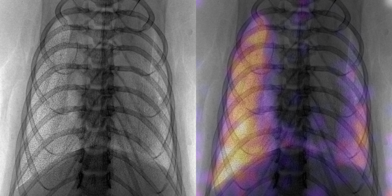
Propagation-based X-ray Phase Contrast (PB-XPC) images of lungs reveal a speckled intensity distribution present only in lung regions. The speckle can be explained as multiple refractions at the numerous air-tissue interfaces the X-rays encounter as they travel through the lung. Information regarding airway microstructure that is encoded within speckle texture of a single XPC radiograph can be decoded to spatially resolve changes in lung properties such as microstructure sizes, air volumes, and compliance. Such functional information cannot be derived from conventional lung radiography or any other 2D imaging modality. By computing these images at different time points within a breathing cycle, dynamic functional imaging can be potentially achieved without the need for tomography. Previously only observed in synchrotron studies, we have demonstrated the first small animal XPC lung speckle images acquired with a bench top system in vivo. This imaging technique is well-suited for a wide range of pre-clinical pulmonary studies which investigate lung function, disease progression, and efficacy of treatment options. Our research collaborators on this project include Dr. Steve Brody and Dr. Buck Rogers at WashU.
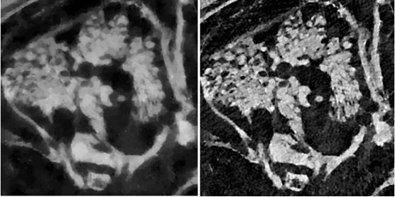
Development of XPC Imaging Technologies for Tissue Engineering and Biomaterials Monitoring
We are developing and evaluating a high-resolution X-ray phase- contrast (XPC) imaging system and associated image reconstruction algorithms for in-vivo volumetric imaging of biomaterials in small animal models. The need for improved imaging methods for evaluating and monitoring biomaterials for tissue engineering/regeneration, drug delivery, and cell therapies applications is great. The ideal method would provide 3D quantitative information, possess high spatial resolution (<100 μm), allow deep tissue penetration (>5 cm), and provide contrast between tissue and material structures essential for evaluating tissue response and development. Currently available imaging methods fall short in one or more of these requirements and this is currently limiting the development of biomaterial-based therapies. Moreover, these limitations hinder a variety of other preclinical imaging applications.
Our research directly addresses the current limitations of high-resolution XPC imaging and will permit its translation for in-vivo volumetric imaging of biomaterials in small animal models. Our approach involves a high degree of innovation regarding both the hardware implementation and image reconstruction methods. Our collaborator on this project is Dr. Eric Brey at UT San Antonio.
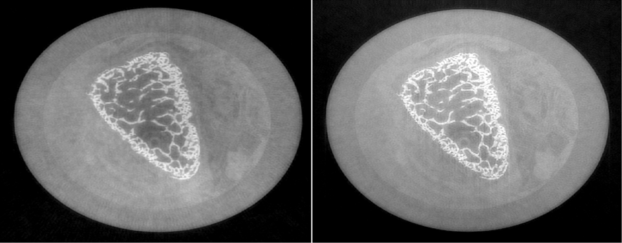
Development of Advanced Image Reconstruction Methods for XPC Tomography
Few-view image reconstruction: An important application of differential X-ray phase-contrast tomography (D-XPCT) within the reach of currently available imaging hardware is high-resolution imaging of biomedical samples. However, reconstructing high resolution D-XPCT images from few-view tomographic measurements remains a challenging task that has not been systematically explored. We have developed a non-conventional objective function used for few-view iterative D-XPCT image reconstruction. It can potentially mitigate the high-frequency information loss caused by data incompleteness and produce images that have better preserved high spatial frequency content than those produced by use of a conventional penalized least squares (PLS) estimator. The proposed algorithm was investigated by use of experimental data produced by an edge-illumination XPCT imager.
Joint image reconstruction methods: Edge illumination X-ray phase-contrast tomography (EIXPCT) is an emerging imaging technique that seeks to circumvent the limitations of previous benchtop implementations of XPC tomography. The goal of EIXPCT is to produce images that separately depict the spatially variant X-ray refractive index and absorption distributions within an object. As with grating- or analyzer-based methods, conventional image reconstruction methods for EIXPCT require that two or more images are acquired at each tomographic view angle. This requirement leads to increased data-acquisition times and radiation doses, which can hinder in vivo applications. To circumvent this, we have developed a joint reconstruction (JR) method that concurrently produces estimates of the refractive index and absorption distributions from a tomographic data set containing only a single image per tomographic view angle. The JR reconstruction method solves a non-linear optimization problem by use of a novel iterative gradient-based algorithms.