We are one of the world’s leading groups pursuing research related to the computational aspects of image formation in PACT. Our research focuses on the development and translation of advanced image reconstruction algorithms or important preclinical and clinical applications. Ongoing projects in our lab include the following:
Advanced Image Reconstruction Development
We are actively engaged in the development of advanced tomographic image reconstruction methods for PACT. This work includes the development of image formation models that include the response of the transducer and other relevant imaging physics. We are also developing robust reconstruction algorithms that can compensate for acoustic heterogeneities within an object mitigate artifacts due to incomplete data.
For 3D imaging applications, we are developing computationally efficient iterative image reconstruction approaches that combine rapidly converging algorithms with hardware acceleration via graphics processing units (GPUs). This work is guided by the extensive use of experimental PACT data, in collaboration with Prof. Lihong Wang at Caltech and Dr. Alexander Oraevsky’s team at TomoWave Laboratories.
Movies:
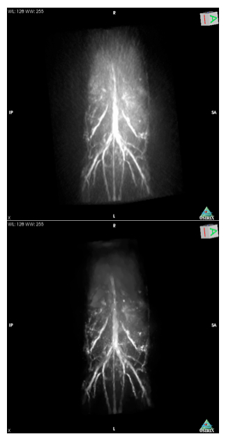
- 3D movie of a mouse reconstructed using filtered back projection algorithm from 180 views
- 3D movie of a mouse reconstructed using advanced iterative algorithm from 90 views
Transcranial PACT Brain imaging
Human brain imaging represents an important imaging application that can benefit tremendously by the development of PACT methods. Existing high-resolution human brain imaging modalities such as X-ray computed tomography (CT) and magnetic resonance imaging (MRI) are expensive and employ bulky and generally non-portable imaging equipment. Moreover, X-ray CT employs ionizing radiation and is unsafe for patients who need long time monitoring of brain diseases or injuries. Alternatively, ultrasonography is an established portable pediatric brain imaging modality, but its image quality degrades severely when employed after the closure of the fontanels and therefore is not effective for imaging adults. The development of PACT brain imaging methods would circumvent these limitations and result a powerful new brain imaging modality that would fill an important void left by the available techniques.
A major technical challenge in PAT brain imaging is to compensate for the distortion introduced into the PACT measurement data by the skull. We are working to make PACT brain imaging a practical, useful, and highly effective brain imaging modality by developing robust image reconstruction methods that can account for skull-induced signal distortion and other physical factors. By use of information regarding the skull morphology and composition obtained from previously acquired adjunct image data, or alternatively from PACT data themselves, we are developing robust imaging models and reconstruction algorithms for PACT that account for the effects of skull-induced wavefront aberrations and other physical factors related to the measurement process. We are developing reconstruction algorithms for obtaining accurate images from incomplete data sets that correspond to clinically useful measurement configurations. This project represents a longstanding collaboration with Professor Lihong Wang at Caltech.
Movies:
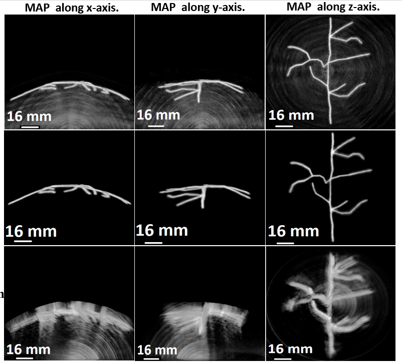
- 3D image obtained after application of the iterative reconstruction algorithm that accounts for shear waves
- 3D image obtained after applying BP reconstruction algorithm that does not account for propagation of shear waves
PACT Breast Imaging
Breast cancer is the second most common cancer type among females in the US. Current treatment of breast cancer still rely on an early detection of tumor, which makes breast imaging a crucial role in the diagnostic process. Existing imaging modalities for breast screening include X-ray mammography, B-mode ultrasound, and breast MRI. X-ray mammography has a high false-positive rate for young females because of the nature of the X-ray contrast; B-mode ultrasound is only used as a supplementary procedure and the result is operator-dependent; breast MRI has high resolution but relatively low contrast, and it is extremely expensive and bulky. PACT, on the other hand, combines the high optical contrast with high ultrasonic spatial resolution. Both the structural and functional information revealed by PACT are valuable for diagnostic purposes. In addition, PACT is radiation- and breast-compression-free. All these advantages make PACT a promising tool for breast imaging.
We have been developing 3D PACT breast imaging systems for clinical use with our collaborators at Tomowave Laborotories and MD Anderson Cancer Center at Houston. A major challenge related to PACT breast imaging is illumination. When performing 3D imaging of large objects such as a female breast, it is difficult to achieve a relatively uniform distribution of light fluence within the volume of interest. To guide hardware designs, we have been investigating different light-delivery and acoustic detecting designs using numerical simulation studies and task-based image quality measures. We are also developing fast, robust, and accurate reconstruction algorithms tailored for PACT breast systems.
Publication
- Seonyeong Park, Frank J. Brooks, Umberto Villa, Richard Su, Mark A. Anastasio, Alexander A. Oraevsky, “Normalization of optical fluence distribution for three-dimensional functional optoacoustic tomography of the breast,” J. Biomed. Opt. 27(3) 036001 (16 March 2022) https://doi.org/10.1117/1.JBO.27.3.036001